
Optomap Ultra Wide-field Retinal Imaging
Optos Wide-Field Retinal Image
The Optomap™ Retinal Exam produces the most comprehensive high-definition wide-field image of your retina, allowing our doctors to perform a more thorough exam. The result is that we can better prevent or reduce vision loss by early detection of eye diseases with simple, quick and patient-friendly technology.
WHY IS OPTOS ULTRA WIDE IMAGING IMPORTANT?
Any routine eye exam should include a careful look at your retina, located at the back of your eye, to assess for abnormalities or disease. The sensitive retinal tissue is susceptible to a variety of conditions, such as diabetes, hypertension, tumors, retinal tears and detachments, macular degeneration, and glaucoma. These conditions can ultimately lead to partial loss of vision or even complete blindness. Early detection of any abnormality is highly desirable. Patients with conditions such as diabetes still need dilation, but this helps the doctor monitor the images year to year.
*IMPORTANT:* The Optomap™ Retinal Scan is an elective procedure that our doctors highly recommend. The fee is $39 for both eyes. This is not billable toward vision plans or medical insurance coverage.
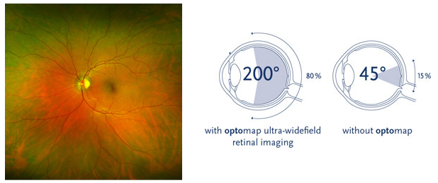
HOW DOES OPTOS IMAGING WORK?
The Optos scan image is safely and comfortably acquired by our technicians with the click of a button using special digital technology in only a quarter of a second. It gives our doctors a high resolution ultra-wide field image. Results will be reviewed with you and will be compared to prior images and current exam room finding.
As can be seen in the two images below, normal versus abnormal is easy to see with the Optos scan.
Normal Retina:
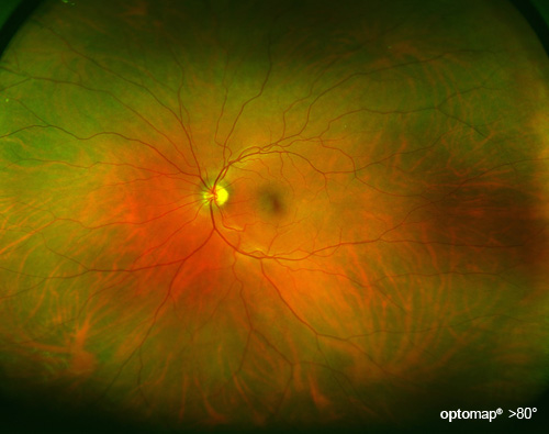
The human retina is about the size of a large postage stamp. This image shows normal blood vessels, optic nerve and retina tissue which is essentially the “wallpaper” on the inside of the eye.
Retina with Lattice Degeneration:
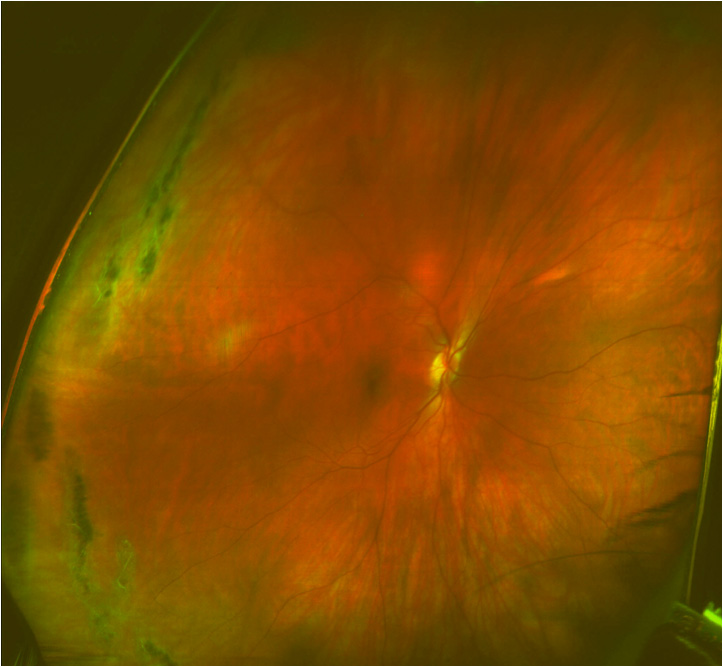
The abnormal areas of this retina are on the left side of this image, looking like stretch marks with dark pigmentation. This condition can lead to retinal holes, tears, or even detachments.