
Optomap Retinal Imaging in Orland Park, IL
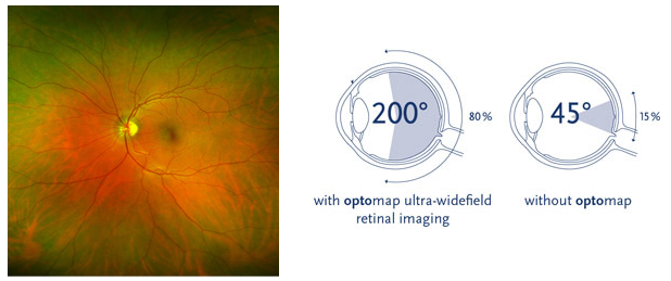
Optomap ultra-widefield retinal imaging provides a broader view of the retina than traditional retinal imaging.
Optomap Retinal Imaging in Orland Park, IL
At Vision Source – Orland Park, we use Optomap ultra-widefield retinal imaging to capture a broad digital image of the retina, the light-sensitive tissue at the back of the eye.
This image helps our doctors evaluate your retinal health, review findings with you during your exam, and compare images over time. Retinal imaging can help detect or monitor signs of diabetes, high blood pressure, retinal tears or detachments, macular degeneration, glaucoma, and other eye health conditions.
Why Retinal Imaging Matters
A careful look at the retina is an important part of a comprehensive eye exam. The retina can show signs of eye disease as well as certain health conditions that affect the blood vessels and tissues inside the eye.
Optomap imaging gives us a wide-field digital view that can help document your eye health and provide a useful baseline for future comparison.
Does Optomap Replace Dilation?
Optomap imaging does not always replace dilation. Patients with diabetes, flashes, floaters, retinal concerns, glaucoma risk, or certain medical findings may still need a dilated eye exam.
Your doctor will recommend the appropriate evaluation based on your symptoms, medical history, and exam findings.
How the Scan Works
The Optomap scan is quick, comfortable, and non-invasive. Our technician captures the image using digital retinal imaging technology, and the doctor reviews the results as part of your examination.
When previous images are available, we can compare them year to year to look for changes.
Normal and Abnormal Retina Examples
Optomap imaging can help patients better understand what the doctor sees during the retinal evaluation. A normal retina shows healthy retinal tissue, blood vessels, and the optic nerve.
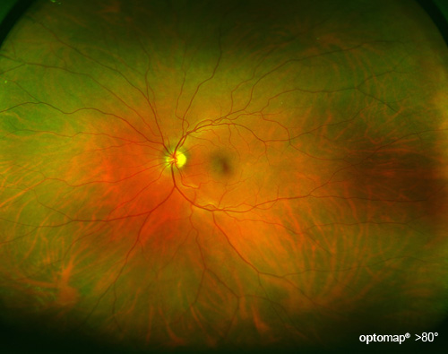
Normal Optomap retinal image showing retinal tissue, blood vessels, and the optic nerve.
In some cases, wide-field imaging may help reveal peripheral retinal findings, such as lattice degeneration, retinal holes, tears, or other areas that may need closer evaluation.
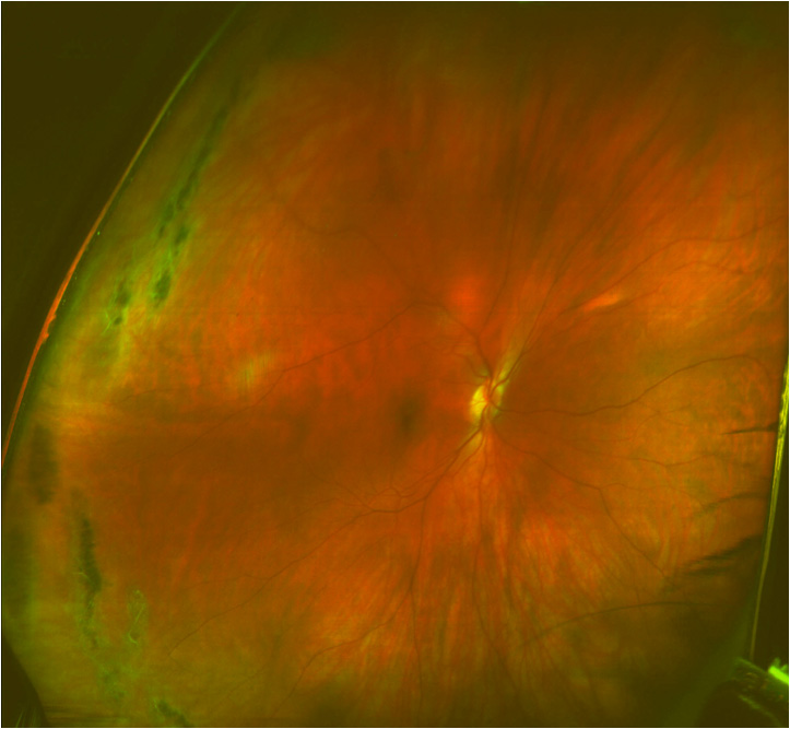
Optomap image showing lattice degeneration, a peripheral retinal finding that may require closer evaluation.
Is There an Additional Fee?
Optomap retinal imaging is an elective procedure that our doctor highly recommends for many patients. The fee is $39 for both eyes. Some vision plans may provide an allowance or reduced copay, but benefits vary by plan.
Schedule a Comprehensive Eye Exam
Optomap retinal imaging may be recommended as part of your comprehensive eye exam. Schedule an appointment at Vision Source – Orland Park to evaluate your vision and eye health.